Liquid embolization is a procedure where small decisions matter: how the catheter is positioned, how stable the distal access is, how the embolic material behaves, how reflux is controlled, and how the operator responds when the anatomy is less forgiving than expected. These skills cannot be developed fully through lectures, videos, or simplified bench demonstrations alone.
For years, many embolization training programs have relied on single-use models, animal labs, cadaveric anatomy, or simplified vessel layouts. These approaches can be useful, but they often limit repeatability, anatomical variation, workflow realism, cost control, and sustainability. Modern neurointerventional training needs a platform that lets physicians repeat the same procedure, compare techniques, evaluate devices, and work across multiple pathologies without rebuilding the entire setup each time.
SurgeonsLab developed its Reusable Liquid Embolic Demonstrator as a practical answer to this need. It is a reusable, fluoroscopy-compatible embolization platform designed for physician training, hands-on workshops, product demonstrations, device evaluation, and patient-specific procedural planning. The same base platform can support interchangeable neurovascular anatomies, including AVM, DAVF, MMA, fistula, tumour embolization, and other endovascular embolization workflows.
Intended use note: This platform is intended for education, simulation, product demonstration, and bench evaluation. It is not intended for direct clinical use.
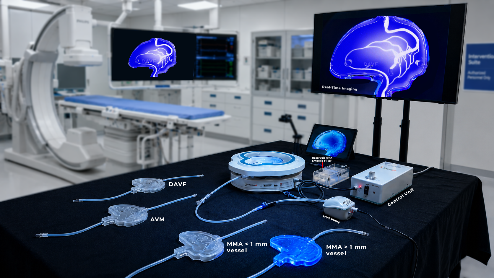
Figure 1. Reusable liquid embolic Demonstrator setup with fluoroscopy-style visualization and interchangeable embolization models.
Why Liquid Embolic Training Needs a Different Kind of Demonstrator
Liquid embolization is not only about delivering an agent. The learning challenge begins much earlier: selecting the access route, shaping the catheter strategy, reaching the target safely, controlling stability during injection, and understanding how the embolic material behaves inside a living-flow environment.
A static model may show anatomy, but it cannot reproduce the training value of flow. A disposable model may support a demonstration, but it can become expensive and wasteful when a training center needs repeated practice across several stations. A generic vascular layout may help with basic catheter handling, but it does not teach the different decision-making required for AVMs, DAVFs, MMA embolization, tumour embolization, or patient-specific cases.
The SurgeonsLab approach is built around three practical requirements: repeatability, realism, and modularity. Trainees can practice the same workflow repeatedly. Instructors can switch anatomies for different procedures. Medical device companies can demonstrate product behavior in a controlled, visible, and reproducible environment.
What Makes the SurgeonsLab Demonstrator Different
The platform combines transparent vascular anatomy, controlled perfusion, fluoroscopy compatibility, and fast model interchangeability. Instead of treating each vascular anatomy as a disposable unit, the system is designed around a reusable base and swappable pathology modules.
- Reusable base platform for repeated training and demonstration sessions.
- Interchangeable anatomy modules for AVM, DAVF, MMA, fistula, tumour embolization, and other neurovascular workflows.
- Fluoroscopy and angio-suite compatibility for hands-on training under image guidance.
- Bench-top demonstration mode for sales training, physician education, and conference workshops.
- Pulsatile and directional flow options to simulate antegrade and retrograde flow conditions.
- Transparent anatomy for direct visual feedback during setup, injection, troubleshooting, and post-run review.
- Patient-specific model development from imaging data when case rehearsal or anatomy-specific demonstration is required.
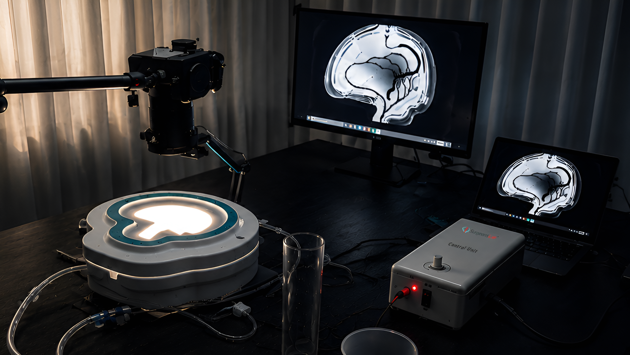
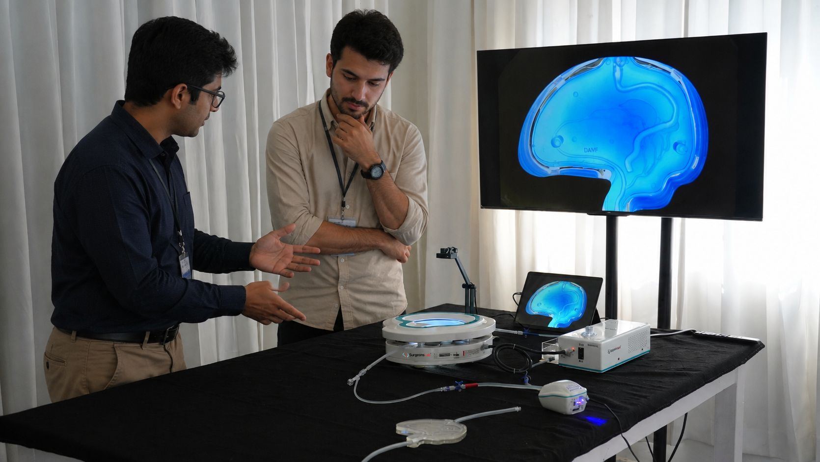
Figure 2. Bench-top reusable Demonstrator configuration with flow, display and control unit for embolization training.
Electromagnetic Fixation: One Part of a Larger System
A key engineering feature of the platform is its magnetic/electromagnetic fixation concept. In embolization simulation, the vessel model must stay aligned, sealed, and stable during flow and device manipulation. Poor fixation creates leaks, movement, inconsistent results, and time-consuming setup.
The fixation approach is designed to hold the model securely while allowing fast exchange between anatomies. This reduces setup time during workshops, protects delicate vascular modules from excessive mechanical clamping, and supports repeatable flow conditions between stations.
Procedural Workflow Training Across Multiple Pathologies
The strongest value of the Demonstrator is not a single feature. It is the ability to reproduce different embolization workflows on one reusable platform. Each pathology requires a different strategy, different device behavior, and different operator decision-making.
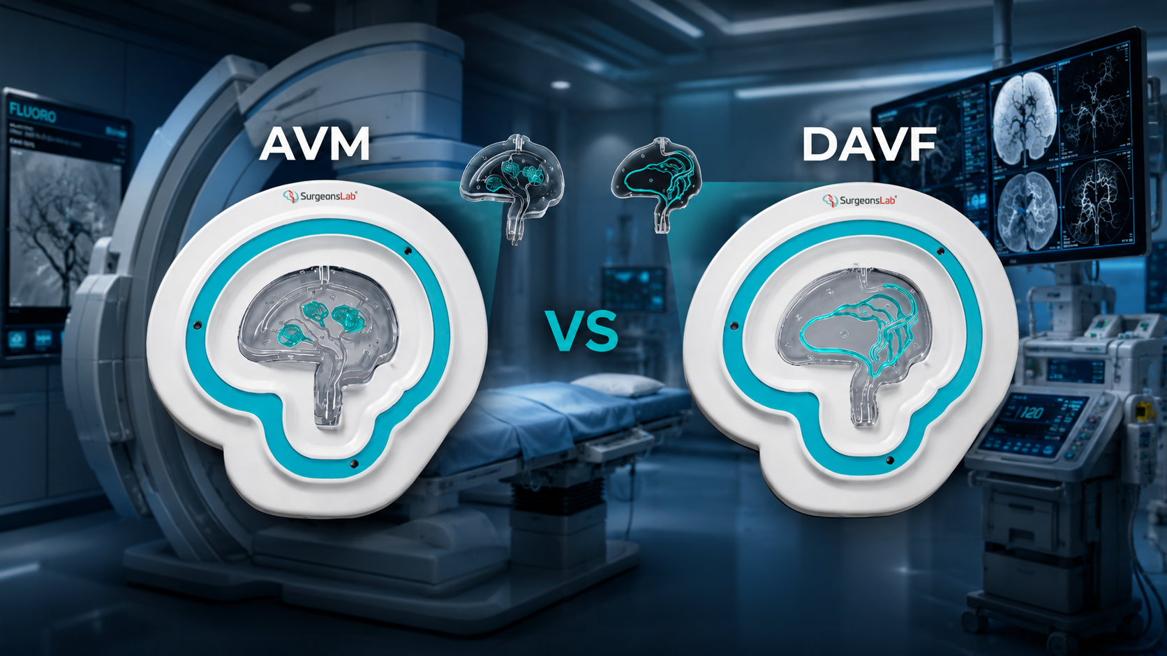
AVM Embolization Training
Arteriovenous malformations require precise microcatheter navigation, careful understanding of feeding arteries, nidus penetration, flow dynamics, reflux control, and staged embolization strategy. SurgeonsLab AVM modules support realistic navigation and embolization training — trainees can practice distal access, microcatheter stability, injection planning, and visual assessment of embolic progression.
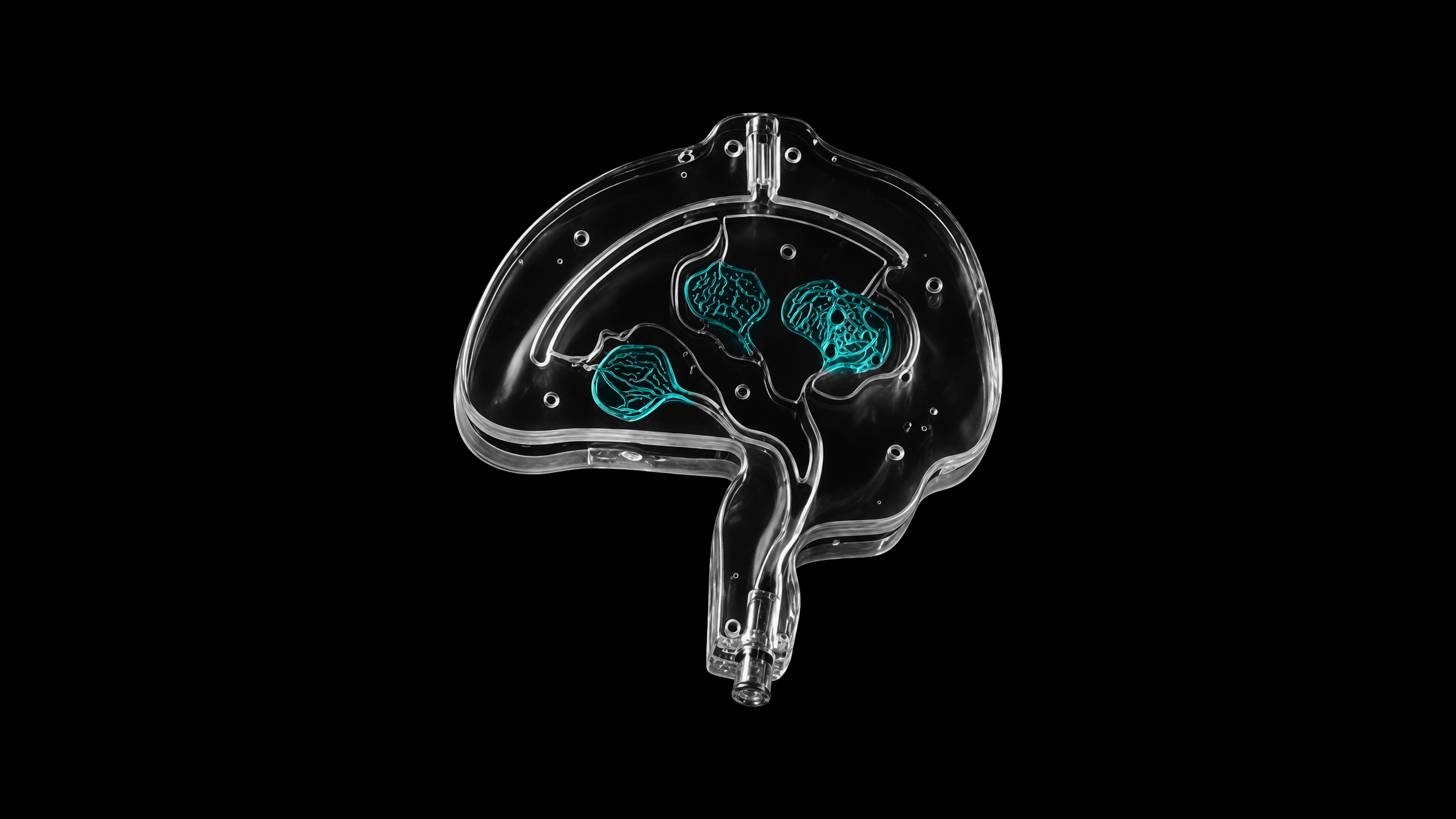
Figure 3. AVM training module with cyan nidus network in a transparent neurovascular model.
DAVF and Fistula Embolization Training
Dural arteriovenous fistulas demand a different mindset from AVM embolization. The operator must identify the fistula point, understand arterial feeders and venous drainage, and decide whether a transarterial, transvenous, or combined strategy fits the training objective. The transparent DAVF and fistula models let instructors show the relationship between feeding vessels, venous pathways, and target zones.
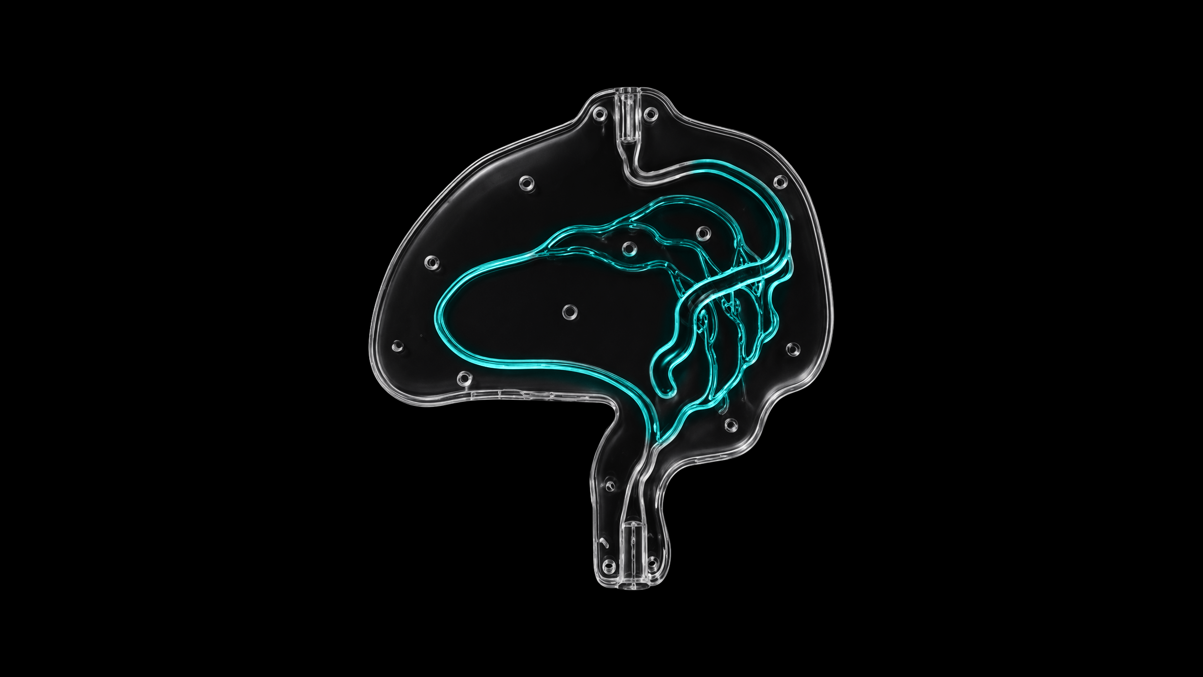
Figure 4. DAVF-style vascular module showing a dural sinus pathway and feeder network.
MMA Embolization Training for Chronic Subdural Hematoma
Middle meningeal artery embolization has become an important, rapidly expanding workflow in neurointervention for chronic subdural hematoma. The focus is often on ECA catheterization, MMA branch selection, distal microcatheter navigation, awareness of dangerous anastomoses, and controlled embolic delivery. SurgeonsLab MMA modules support both 2D and 3D layouts, including vessel sizes above and below 1 mm.
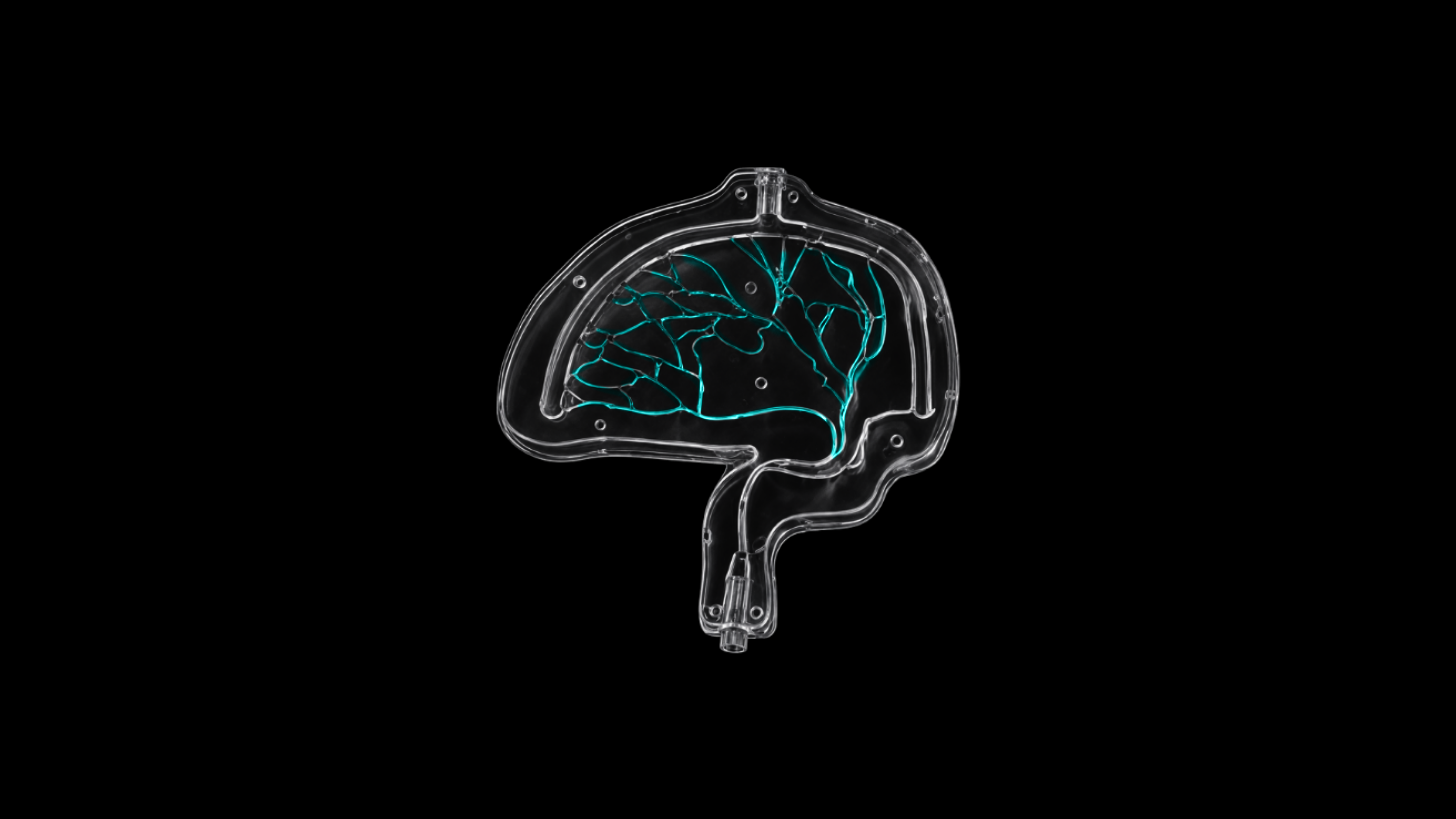
Figure 5. Fine-vessel MMA training module for microcatheter navigation and distal branch selection.
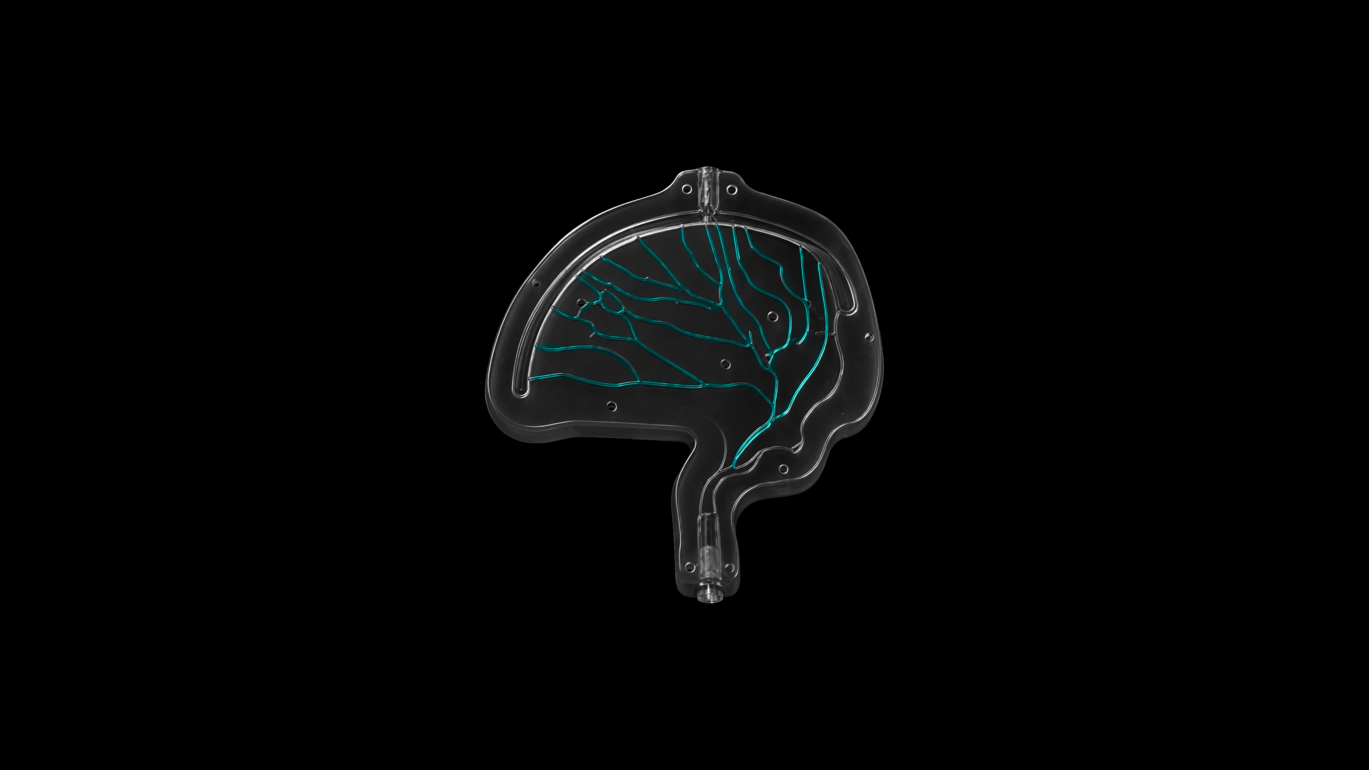
Figure 6. MMA embolization module showing a wider branch network for over-1-mm vessel training.
Tumour Embolization, Small-Vessel Training and Emerging Applications
The same reusable platform can also support tumour embolization, small-vessel embolization, selective catheterization practice, and emerging embolic technology demonstrations. For institutions and companies running frequent courses, this modularity matters — a single infrastructure can support multiple sessions, different skill levels, and different product categories.
Realistic Flow and Material Behavior
Embolization is flow-dependent. A training model should help users understand how injection force, catheter stability, vessel diameter, flow direction, and embolic properties interact. SurgeonsLab models can be paired with controlled perfusion to create pulsatile or directional flow environments, including antegrade and retrograde conditions depending on the module and training objective.
The Demonstrator can be configured for training with common embolization workflows, including DMSO/EVOH-style liquid embolic workflows, NBCA-style techniques, particles, coils, microspheres, and emerging embolization technologies.
Material compatibility and cleaning protocols should be confirmed for each agent, product, and intended use case before a workshop or evaluation program.
Fluoroscopy-Compatible and Bench-Top Friendly
The platform supports both angio-suite use and bench-top demonstration. In a fluoroscopy environment, trainees work under imaging guidance and practice the visual decision-making required in interventional neuroradiology. In a bench-top setup, instructors and device teams can demonstrate anatomy, access strategy, and embolic behavior without the complexity of a full angio lab.
"Resident, fellow, and early-career neurointerventional training built around repeatability, not single-use anatomy."
Patient-Specific Embolization Planning and Case Rehearsal
Generic models are useful for structured training, but patient-specific anatomy can change the conversation completely. SurgeonsLab can support model development from imaging datasets such as CTA, MRA, DSA, and DICOM-based reconstructions — converting complex vascular anatomy into a physical, transparent simulation model for education, planning, discussion, and rehearsal.
For difficult AVMs, DAVFs, unusual MMA anatomy, or complex access routes, a patient-specific model can help the team understand the anatomy before entering the angio suite, supporting pre-procedure discussion, device selection, access planning, and communication across the care team.
Applications for Hospitals, Training Centers and Medical Device Companies
The reusable liquid embolic Demonstrator is designed for multiple stakeholder groups. Each uses the platform differently, but all benefit from repeatability, visibility, and modular anatomy.
Hospitals & Academic Centers
- Resident, fellow, and early-career training
- Structured embolization workshops with repeated stations
- Team training for physicians, nurses, and technologists
- Pre-procedure planning for complex anatomies
- Simulation-based education without animal labs
Medical Device Companies
- Hands-on physician education and KOL workshops
- Product launch demonstrations
- Sales and clinical specialist training
- Bench evaluation under controlled conditions
- Conference booth demonstrations
Economic and Sustainability Advantages of Reusability
High-quality embolization training can become expensive when every session requires a new disposable model. Reusability changes the cost structure — a reusable base platform with interchangeable anatomy modules allows institutions and companies to run more sessions, train more users, and reduce waste per workshop.
This matters especially for large courses, multi-station workshops, international roadshows, device launch programs, and recurring fellowship training.
Why SurgeonsLab
SurgeonsLab is focused on humanized surgical and endovascular simulation: transparent anatomy, realistic workflows, patient-specific model development, and practical training systems that work with real instruments and imaging environments. The reusable liquid embolic Demonstrator fits within the broader SurgeonsLab ecosystem of CathTrain endovascular simulation, patient-specific flow models, and procedure-focused training platforms.
How a Typical Workshop Can Be Structured
A reusable liquid embolic workshop can be designed around skill progression — anatomy orientation, microcatheter navigation, injection strategy, and post-embolization review. The same platform can then be reset for the next user or switched to a different pathology.
- Station 1: Anatomy orientation and access pathway planning
- Station 2: Microcatheter and microwire navigation to target vessels
- Station 3: AVM nidus embolization workflow and reflux control
- Station 4: DAVF/fistula target identification and embolization strategy
- Station 5: MMA branch selection and distal embolization workflow
- Station 6: Device demonstration, product comparison, or patient-specific case discussion
Conclusion
Liquid embolization training needs to move beyond single-use anatomy and generic demonstrations. Physicians need repeatable procedural practice. Training centers need scalable platforms. Device companies need visible, realistic, and reproducible environments for education and product demonstration.
The SurgeonsLab Reusable Liquid Embolic Demonstrator brings these needs together in one modular platform — supporting AVM, DAVF, MMA, fistula, tumour embolization, patient-specific planning, fluoroscopy-compatible training, and bench-top demonstration.
Request a Demonstration
SurgeonsLab can support hands-on workshops, institutional training programs, conference demonstrations, product evaluation sessions, and custom pathology development.
Request a demonstration Contact SurgeonsLabFrequently Asked Questions
What procedures can be trained on the reusable liquid embolic Demonstrator?
The platform can support AVM embolization, DAVF and fistula embolization, MMA embolization for chronic subdural hematoma, tumour embolization, small-vessel catheterization, and other configurable neurovascular embolization workflows.
Is the Demonstrator compatible with fluoroscopy?
Yes. The models are designed for fluoroscopy-compatible and angio-compatible training, while also supporting bench-top demonstration when a full imaging suite is not required.
Can the same base platform be used for different anatomies?
Yes. The reusable base is designed to support interchangeable pathology modules, allowing one platform to be used across different procedures, user levels, and workshop stations.
Can the Demonstrator support DMSO/EVOH or NBCA-style workflows?
The Demonstrator can be configured for selected liquid embolic workflows, including DMSO/EVOH-style and NBCA-style training. Material compatibility, cleaning protocols, and intended use should be confirmed for each agent or product before use.
Does SurgeonsLab create patient-specific embolization models?
Yes. SurgeonsLab can develop patient-specific models from imaging datasets such as CTA, MRA, DSA, and DICOM reconstructions when case-specific planning, education, or demonstration is needed.
Who benefits most from this platform?
Neurointerventionalists, neurosurgeons, interventional radiologists, fellows, training centers, hospitals, medical device companies, sales teams, clinical specialists, and KOL-led education programs.
How does reusability reduce cost?
A reusable base platform reduces the need to purchase or dispose of a complete new model for every session. Interchangeable anatomy modules allow repeated use across courses, workshops, roadshows, and product demonstrations.