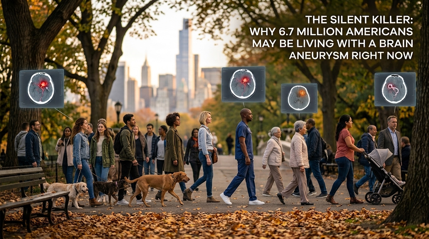
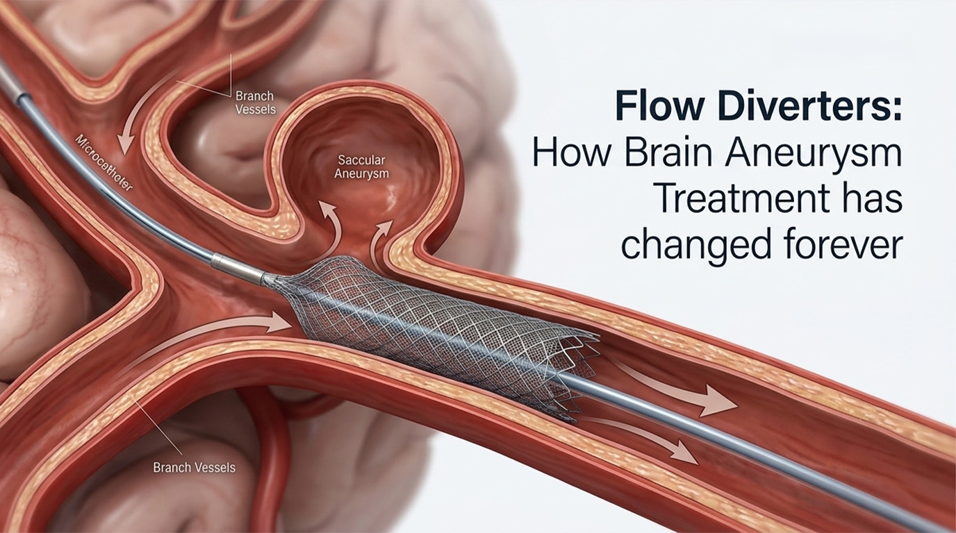
Precision medicine spent two decades reshaping oncology and cardiology. It is now picking up pace in neurovascular care as well. This shift is particularly visible in the treatment of intracranial aneurysms.
Reading Rupture Risk From the Aneurysm Wall Itself
The diagnostic side of this story is advancing at the University of Iowa, where researchers are focusing on developing biologically validated imaging biomarkers for the non-invasive assessment of intracranial aneurysm wall instability. The work builds on years of prior research at Iowa into aneurysm wall enhancement (AWE). AWE is based on the idea that contrast uptake in an aneurysm's wall on high-resolution MRI correlates with how unstable — and therefore how susceptible to rupture — the aneurysm actually is.
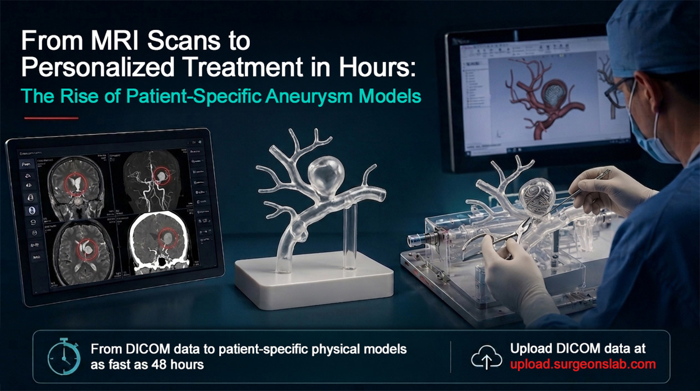
Figure 1. Orthogonal probes extending from the aneurysm lumen into the wall map contrast enhancement in three dimensions, flagging focal regions linked to instability.
Iowa studies had earlier mapped AWE in three dimensions using orthogonal probes extending from the aneurysm lumen into the wall, producing heat-maps that flagged focal regions of enhancement in aneurysms that had ruptured or caused symptoms. The newly funded research work pushes that work toward biological validation, tying the imaging signal to the underlying tissue biology, so that a clinician can look at a single high-res MRI and get a real answer to the question every neurosurgeon and interventionalist asks first:
"Is this particular aneurysm likely to rupture?"
Earlier, this question used to take years of population-level data and a fair amount of clinical judgment to answer with confidence. Increasingly, it can now be answered from advanced imaging and structural mapping.
Acting on the Risk: Patient-Specific Simulation
Identifying the risk is only one half of individualized care. The other half is for the physician to be able to act on it. This is where patient-specific simulation models are plugging the gap.
Patient-specific models such as the ones made by SurgeonsLab are structural and functional replicas of the patient's anatomy. The same imaging data from a CT, MRI, or DSA that is used for diagnosis is converted into a true-to-scale, 3D-printed replica of that patient's exact vasculature and aneurysm. The model has the same tortuosity, same wall thickness, and same sac geometry as the patient's anatomy, and also runs under pulsatile flow that mimics real hemodynamics.
Now, the endovascular team can rehearse a generic clipping or even a coiling technique on the patient's exact anatomy with real instruments, real devices, and real complications built into the personalized model. The team can plan device selection, clip trajectory, and catheter approach on a physical twin of the patient's anatomy — not rely on the statistical average of similar cases.
A 48-Hour Window That Changes the Workflow
Put these two advances together and the resulting workflow looks genuinely new. Earlier, similar anatomical simulation modeling took weeks of manual segmentation and fabrication. Now, SurgeonsLab can deliver patient-specific models in as fast as under 48 hours. This matters enormously because when the biomarker research is telling you that the aneurysm in question is unstable now, the interventional team has a narrow window before a rupture occurs, adding new complications to the procedure.
This article is intended for general education purposes only and does not provide medical advice or replace medical evaluation and individualized clinical advice.
References
SurgeonsLab's educational articles are based on evidence-backed information and reviewed against published clinical and regulatory sources to help ensure accuracy and up-to-date reliability.
View Sources
- Brain Aneurysm Foundation Announces its 2026 Research Grant Recipients, PR Newswire. www.aol.com/articles/brain-aneurysm-foundation-announces-2026-120000000.html
- Stroke: Vascular and Interventional Neurology — Topographical Analysis of Aneurysm Wall Enhancement With 3-Dimensional Mapping. www.ahajournals.org/doi/10.1161/SVIN.121.000309
- Gudino, Andres et al. “E-174 Three-dimensional aneurysm wall enhancement as an imaging biomarker to detect the symptomatic aneurysm in patients with multiple intracranial aneurysms.” Journal of NeuroInterventional Surgery 17 (2025): A210-A211. doi:10.1136/jnis-2025-SNIS.289. www.researchgate.net/publication/393678134
- Raghuram, Ashrita et al. “3D aneurysm wall enhancement is associated with symptomatic presentation.” Journal of Neurointerventional Surgery vol. 15,8 (2023): 747-752. doi:10.1136/jnis-2022-019125. pubmed.ncbi.nlm.nih.gov/35853699
- Raghuram, Ashrita et al. “Abstract 1122‐000213: Presence of Blebs Associated with Reduced Aneurysm Wall Tension and Increased Wall Enhancement.” Stroke: Vascular and Interventional Neurology vol. 1, Suppl 1 e12120. 22 Dec. 2021, doi:10.1161/SVIN.01.suppl_1.000213. pmc.ncbi.nlm.nih.gov/articles/PMC12772841
- Aneurysms, training flow models & neurosurgery simulation models by SurgeonsLab. www.surgeonslab.com
See Your Case as a Patient-Specific Model
If your clinical team or device team wants to see how your imaging will look as a patient-specific model, upload the DICOM data of the case directly and schedule a call with us.
Upload DICOM data Contact SurgeonsLabFrequently Asked Questions
What is aneurysm wall enhancement (AWE)?
AWE is an imaging finding on high-resolution MRI where contrast uptake in an aneurysm's wall correlates with how unstable, and therefore how susceptible to rupture, the aneurysm is.
How is AWE mapped in three dimensions?
Researchers at the University of Iowa have used orthogonal probes extending from the aneurysm lumen into the wall to produce heat-maps that flag focal regions of enhancement, particularly in aneurysms that had ruptured or caused symptoms.
What is a patient-specific aneurysm model?
It is a true-to-scale, 3D-printed replica of a patient's exact vasculature and aneurysm, built from that patient's own CT, MRI, or DSA imaging data. It reproduces the same tortuosity, wall thickness, and sac geometry, and can run under pulsatile flow that mimics real hemodynamics.
How is a patient-specific model different from a generic training model?
A generic model reflects a statistical average of similar cases. A patient-specific model reflects one patient's exact anatomy, allowing the team to rehearse clipping or coiling and plan device selection, clip trajectory, and catheter approach on a physical twin of that specific case.
How quickly can a patient-specific model be produced?
SurgeonsLab can deliver patient-specific models in as fast as under 48 hours, compared to the weeks that manual segmentation and fabrication used to take. This turnaround matters when imaging suggests an aneurysm is unstable and the interventional team has a narrow window to plan before rupture risk increases.
How can a clinical or device team request a patient-specific model?
Teams can upload the DICOM data for a case directly at upload.surgeonslab.com and schedule a call with SurgeonsLab to discuss the resulting model.