A brain aneurysm can be asymptomatic for years. You may never have a headache or any obvious warning sign that an area of a cerebral artery is weak — and that is what makes the numbers striking.
One in every 50 Americans is living with an intact aneurysm. That is an estimated 6.7 million people in the United States who are walking around with an unruptured brain aneurysm.
What Is a Brain Aneurysm?
A cerebral aneurysm, also called an intracranial aneurysm, is a bulging or ballooning area in a blood vessel in the brain.
An unruptured aneurysm may be discovered incidentally during imaging performed for another reason, or it may be identified because a patient has symptoms or risk factors such as smoking, hypertension, and family history that prompt vascular imaging.
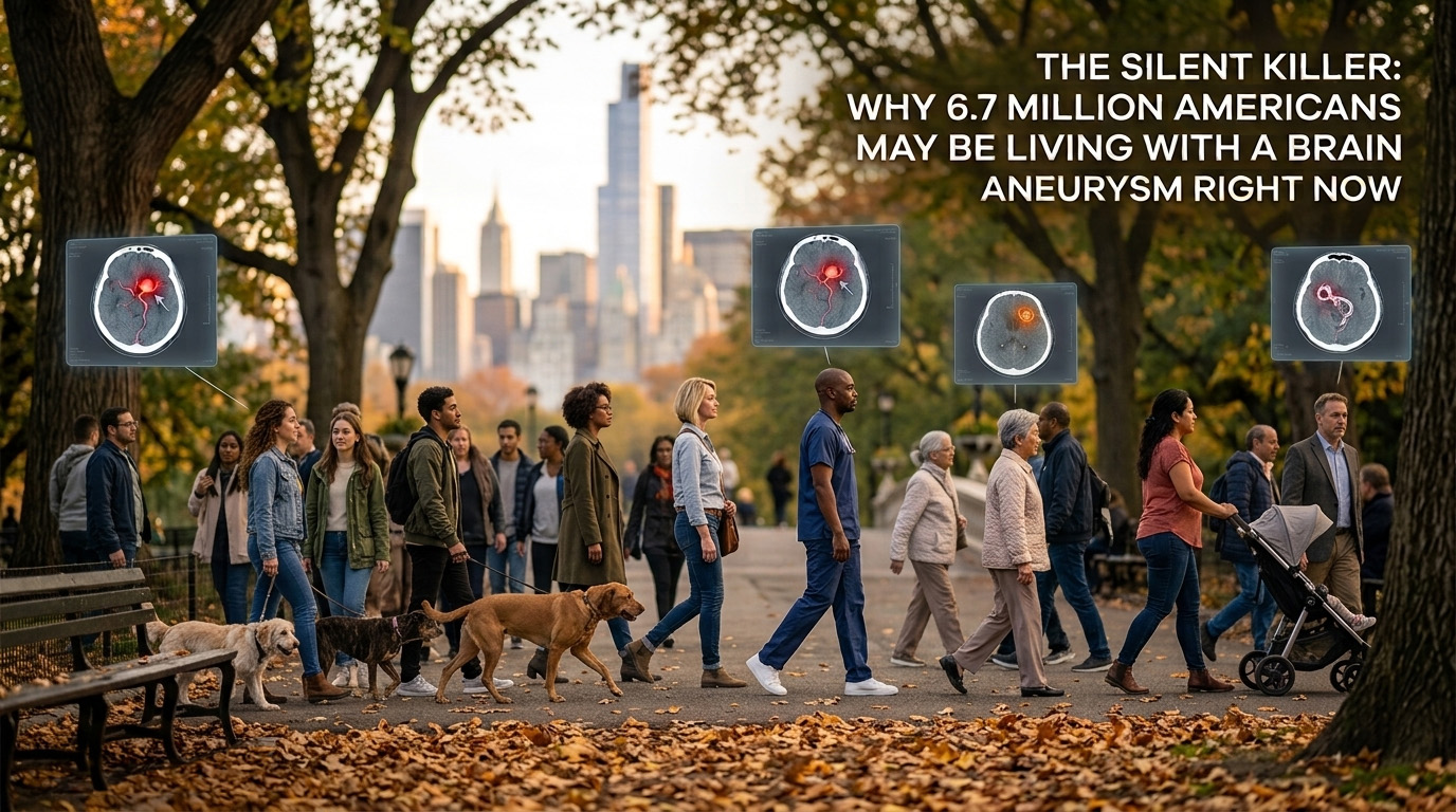
Figure 1. A cerebral aneurysm forms as a weakened, balloon-like bulge in the wall of a brain blood vessel.
The good thing is most people never experience an aneurysm rupture. But in the off-chance that they do, it can cause subarachnoid hemorrhage, a life-threatening neurological emergency. The classic warning is a sudden, severe “Thunderclap” headache, often described as the “Worst Headache” of a person’s life.
The reassuring part is that an unruptured brain aneurysm is not a medical emergency. The physician will assess the size, location, morphology, growth, and the patient’s individual risk factors before recommending either monitoring the aneurysm, an interventional procedure, or any of the other management strategies.
How Are Brain Aneurysms Detected?
Modern neurovascular imaging gives physicians several ways to investigate suspected aneurysms. CT angiography (CTA), magnetic resonance angiography (MRA), and digital subtraction angiography (DSA) can provide detailed views of cerebral blood vessels. But interpreting these images can be demanding, particularly when aneurysms are small, anatomy is complex, or imaging workloads are high.
AI-based aneurysm detection systems can analyze medical images such as CT angiography (CTA) and help flag scans containing suspected intracranial aneurysms. In the U.S., these systems have been cleared by the FDA and are supporting clinical workflows to flag and prioritize suspected saccular aneurysms on head CT.
"AI can assist with detection, while specialist interpretation is still essential to interpret the original imaging, understand the patient's clinical context, and determine what happens next."
This technology is promising. A systematic review and meta-analysis found that AI demonstrated strong potential for cerebral aneurysm detection, but also highlighted substantial limitations including retrospective study designs and concerns about generalizability. More recent clinical research similarly shows that AI can assist with detection, while specialist interpretation is still essential to interpret the original imaging, understand the patient's clinical context, and determine what happens next.
For millions of Americans who may be living with an aneurysm, this combination of advanced imaging technology, clinical expertise, and awareness could be significant.
This article is intended for general education purposes only and does not provide medical advice or replace medical evaluation and individualized clinical advice.
References
SurgeonsLab's educational articles are based on evidence-backed information and reviewed against published clinical and regulatory sources to help ensure accuracy and up-to-date reliability.
View Sources
- Mayo Clinic Minute: The difference in brain aneurysms – Mayo Clinic News Network. newsnetwork.mayoclinic.org/discussion/mayo-clinic-minute-the-difference-in-brain-aneurysms
- Rapid Aneurysm Triage and Notification, FDA 510(k) Summary. www.accessdata.fda.gov/cdrh_docs/pdf23/K230074.pdf
- Din, Munaib et al. “Detection of cerebral aneurysms using artificial intelligence: a systematic review and meta-analysis.” Journal of Neurointerventional Surgery vol. 15,3 (2023): 262-271. doi:10.1136/jnis-2022-019456. pubmed.ncbi.nlm.nih.gov/36375834
- Akinpelu, Babatunde J et al. “Assessing the Accuracy of Artificial Intelligence in Detecting Intracranial Aneurysms in a Clinical Setting Relative to Neuroradiologists.” AJNR. American Journal of Neuroradiology vol. 46,12 2545-2552. 4 Dec. 2025, doi:10.3174/ajnr.A8897. pmc.ncbi.nlm.nih.gov/articles/PMC12687988
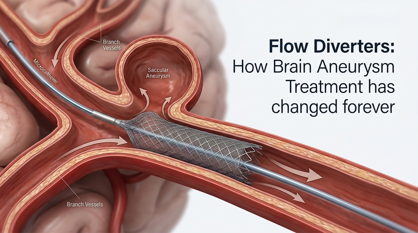
Training for Aneurysm Embolization & Flow Diversion
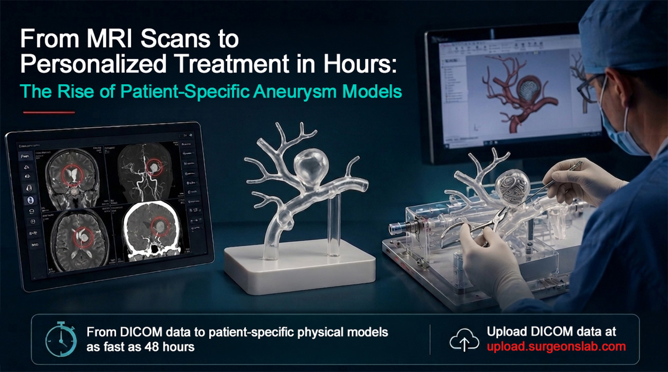
SurgeonsLab develops reusable, fluoroscopy-compatible neurovascular simulation platforms — including patient-specific aneurysm models — for physician training, device evaluation, and procedural planning.
Request a demonstration Contact SurgeonsLabFrequently Asked Questions
How common are unruptured brain aneurysms?
An estimated 1 in every 50 Americans roughly 6.7 million people is living with an intact, unruptured brain aneurysm.
Does having a brain aneurysm mean it will rupture?
No. Most people with an unruptured aneurysm never experience a rupture. Physicians assess size, location, morphology, growth, and individual risk factors before deciding between monitoring, an interventional procedure, or another management strategy.
How are brain aneurysms typically found?
Aneurysms are often found incidentally during imaging performed for another reason, or through CT angiography (CTA), MR angiography (MRA), or digital subtraction angiography (DSA) ordered due to symptoms or risk factors like smoking, hypertension, or family history.
What role does AI play in aneurysm detection?
FDA-cleared AI systems can analyze CTA scans and flag suspected intracranial aneurysms to help prioritize cases for specialist review. Research shows strong potential, but specialist interpretation remains essential for final diagnosis and clinical decision-making.
What is a "thunderclap" headache?
A thunderclap headache is a sudden, severe headache, often described as the worst headache of a person's life. It is a classic warning sign of a ruptured aneurysm and subarachnoid hemorrhage, which is a life-threatening neurological emergency requiring immediate medical attention.